

Understanding the cellular ecosystems that shape human disease
We are a systems biology and precision medicine research group investigating how cellular interactions, molecular signalling, and tissue architecture shape cancer biology and therapeutic response.


Latest News
“To provide a more integrated approach to informing future precision medicine strategies, we combined the complementary strengths of multiple technological platforms to profile multi-regional, spatially annotated surgical biospecimens from 65 RCC patients by single-cell RNA sequencing with paired TCR and BCR repertoire analysis, imaging mass cytometry, suspension mass cytometry, spatial transcriptomics and deconvolved bulk RNA sequencing. With this resource dataset, we explored patient subgroups and precision immunotherapy strategies using an integrated analysis of transcripts and proteins across single cell and spatial modalities.”
Read more at:
https://doi.org/10.64898/2026.08.17.744644
Read more of our archived news updates and stay up to date by following us on Twitter.
Our Research
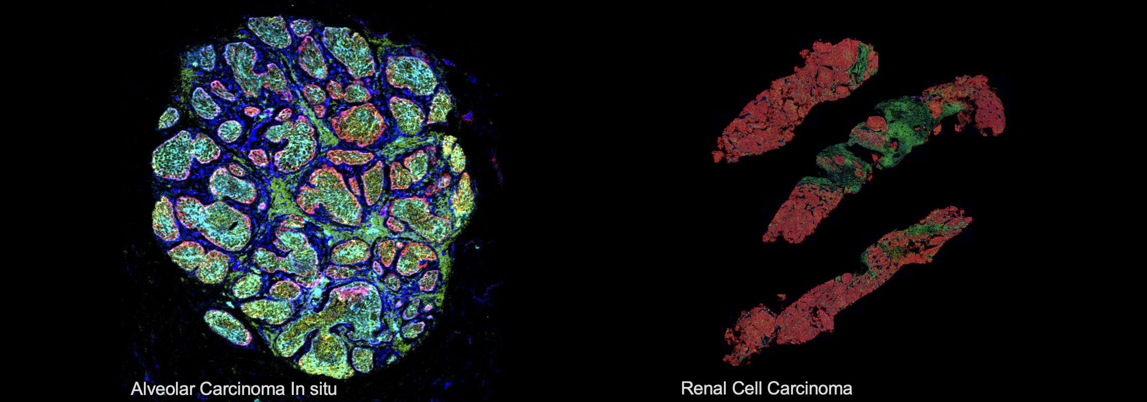
To understand tissues and tumours as integrated systems shaped by their individual cellular components, we use multiplexed imaging technologies to simultaneously quantify single-cell phenotypes, functional states, and cellular interactions, as well as their organization and contribution to tissue architecture. By integrating spatially resolved measurements of molecular and cellular networks across multiple biological scales—from subcellular features to individual cells and organized tissues—we aim to uncover how these complex interactions contribute to health and disease. Our work focuses on quantitative, spatially resolved multiplexed technologies, particularly Imaging Mass Cytometry (IMC), which enable us to perform spatially resolved screening of distinct clones in mouse models of disease and identify biomarkers associated with clinical outcomes in biobanked patient tissues.


Translational Research and Precision Medicine
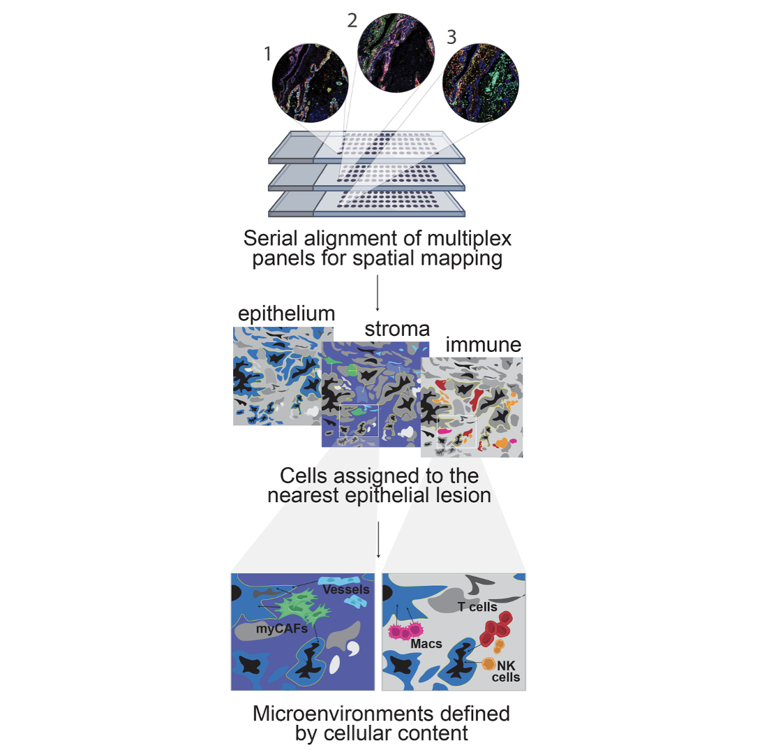
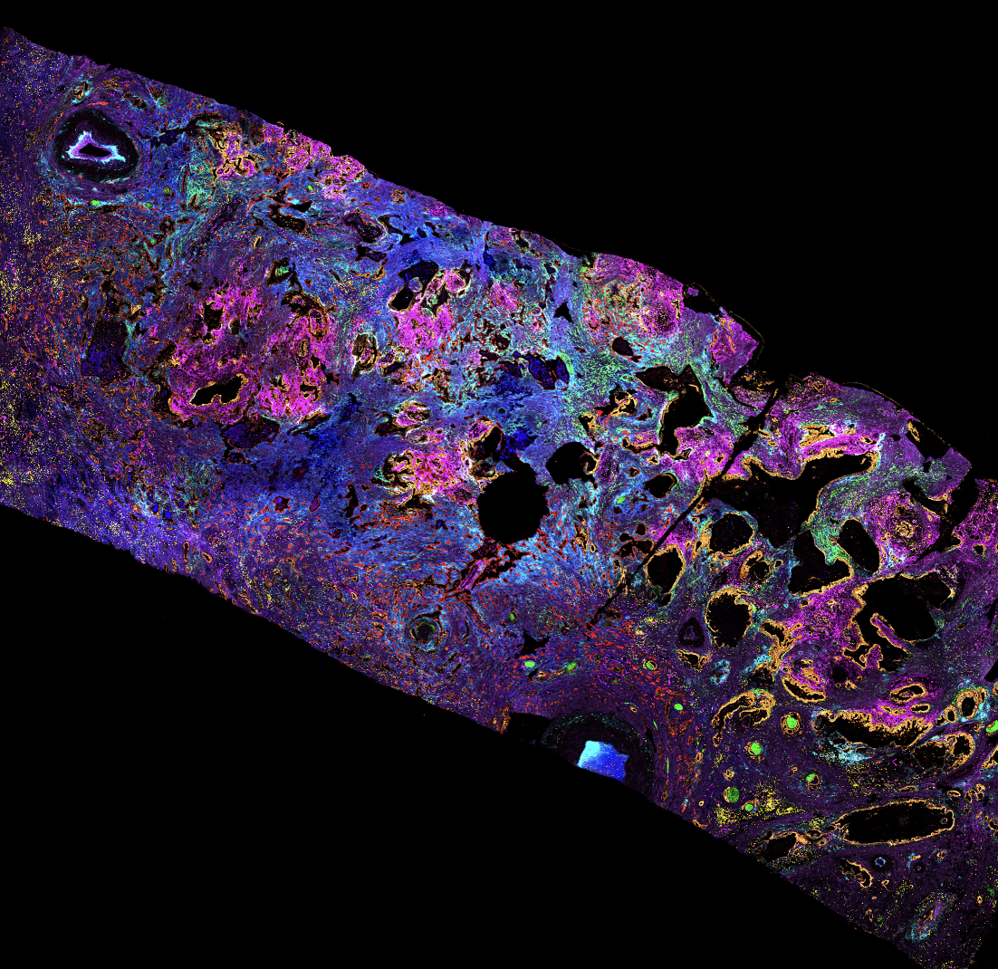
As part of the Pancreatic Cancer Translational Research Initiative (PanCuRx) at the Ontario Institute of Cancer Research, we are studying how intra- and inter-tumour cellular heterogeneity contributes to therapeutic resistance and poor outcomes in Pancreatic Ductal AdenoCarcinoma. By leveraging serial tissue sections from a large patient cohort, we combine high-dimensional single-cell profiling with spatial analysis to characterize cellular phenotypes, functional states, and cell–cell interactions, while uncovering how these features shape the organization and architecture of the tumour microenvironment. This integrated approach allows us to identify complex multi-cellular networks that may inform more effective and personalized clinical decision making.
Mechanisms of Tumour Initiation and Progression
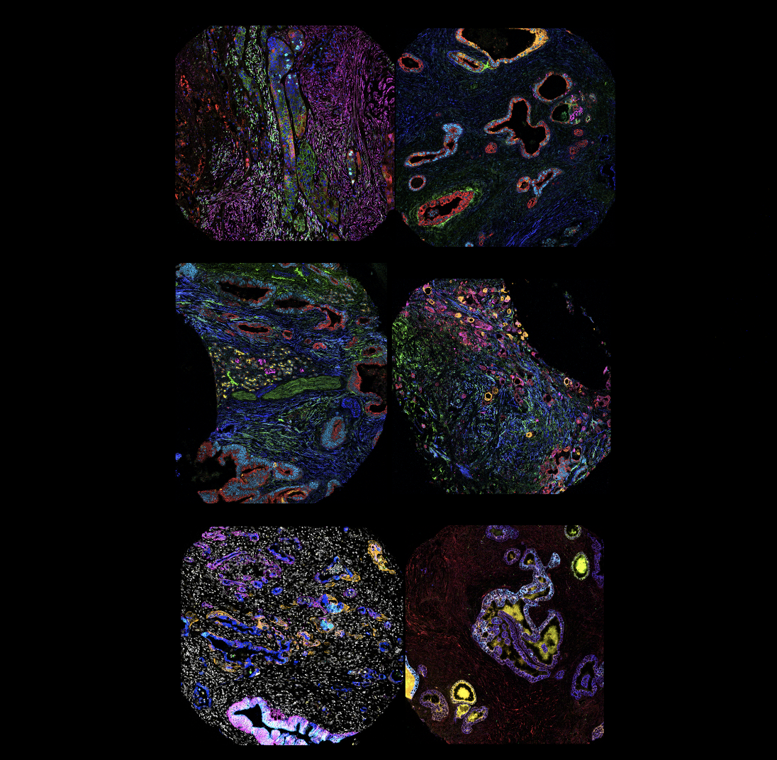
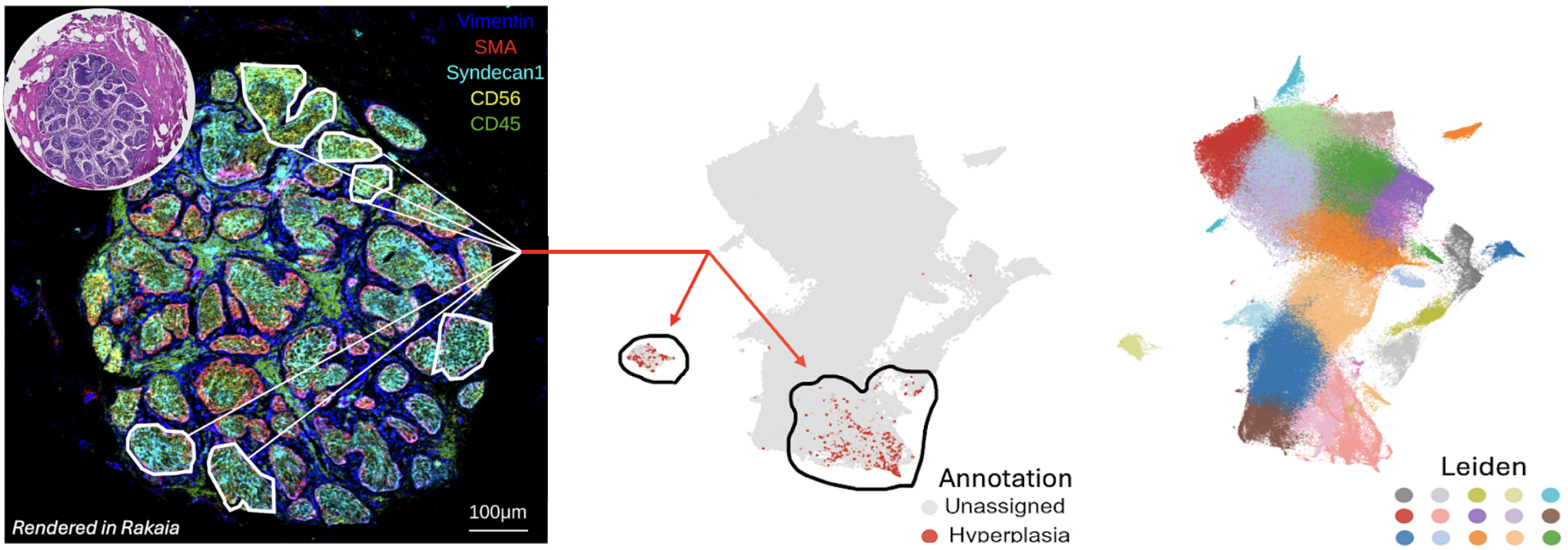
By integrating mouse models of mammary tumorigenesis with highly multiplexed cellular profiling of human breast cancers, we investigate the molecular pathways that drive breast cancer initiation and metastasis. In particular, we are interested in understanding how changes in tumour cell states, interactions with surrounding stromal and immune populations, and alterations in tissue organization contribute to the earliest stages of cancer development and the acquisition of metastatic potential.
This work is made possible through close collaborations with researchers at the Lunenfeld-Tanenbaum Research Institute and clinical teams at Mount Sinai Hospital, who have established the Breast Translational Research Resource. Additionally, we are part of the Terry Fox Research Institute Hippo Pathway Team, and through this collaboration we have developed a spatially resolved functional genomics screening platform to dissect the extracellular signalling networks regulated by tumour-specific Hippo pathway activity.
Spatial Biology Across Disease Contexts
We apply spatially resolved systems biology approaches across diverse disease contexts to investigate how cellular heterogeneity and interactions shape tissue architecture and contribute to disease progression. By integrating high-dimensional multiplexed cellular profiling with quantitative spatial analysis, we characterize cellular phenotypes, functional states, and multicellular organization to uncover the biological mechanisms underlying disease and therapeutic response. Our work spans both the retrospective analysis of large biobanked patient cohorts and the prospective study of small biopsy specimens, each presenting distinct technical and analytical challenges. Ultimately, we aim to identify disease-associated cell states and multicellular networks that can inform clinical decision-making and reveal new opportunities for therapeutic intervention
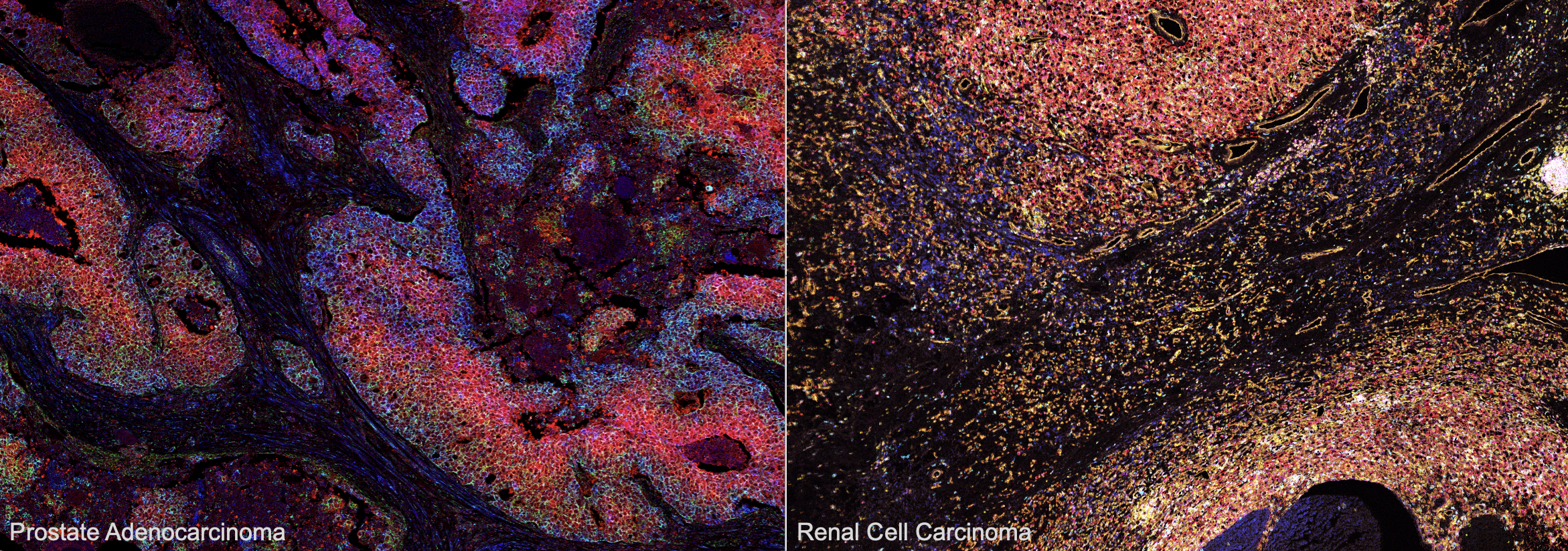
For example, through a US Department of Defense-funded collaboration with urology surgical teams at Toronto General Hospital and Memorial Sloan Kettering Cancer Centre, we are investigating how the extent, composition, and spatial organization of immune cell populations vary across patient tumours. In particular, we aim to understand how the organization of immune cell communities within the tumour microenvironment influences anti-tumour immunity and response to immunotherapy.
Methods Development and Technological Innovation
We develop spatially resolved systems biology tools and analytical approaches to characterize complex tissue environments across multiple biological scales—from genomic and pathway-specific signalling to cellular phenotypes and tissue-level organization. With specific expertise in Imaging Mass Cytometry and CyTOF technology, we integrate spatially resolved ‘omics’ approaches to investigate the molecular mechanisms underlying histopathological features and uncover the complexity of tumour morphology, composition, and architecture. As part of the Medicine by Design Grand Challenges program we are working with a multidisciplinary team of synthetic biologists, chemists, bioengineers, and computational biologists to engineer scalable multi-cellular environments.
Our work is driven by the continuous development of quantitative analytical methods, novel reagents to measure cellular function, and innovative functional and phenotypic screening approaches. By pushing the boundaries of spatial biology, we aim to deepen our understanding of complex tissue systems and uncover new biological insights.
Novel Functional and Phenotypic Screening Approaches
By utilizing a combination of genetic tools with spatially resolved quantitative single-cell approaches in vivo, we investigate the development of cancer, from the initial cells that give rise to tumours through localized sites of dissemination and the formation of distant metastases. Through these approaches, we aim to understand how early changes in cellular states and interactions shape tumour development, and are working to target the molecular bottlenecks that mitigate advanced disease. Explore, interrogate, and prioritize features of interest across large-scale multiplexed imaging datasets
Quantitative Spatial Analysis tools
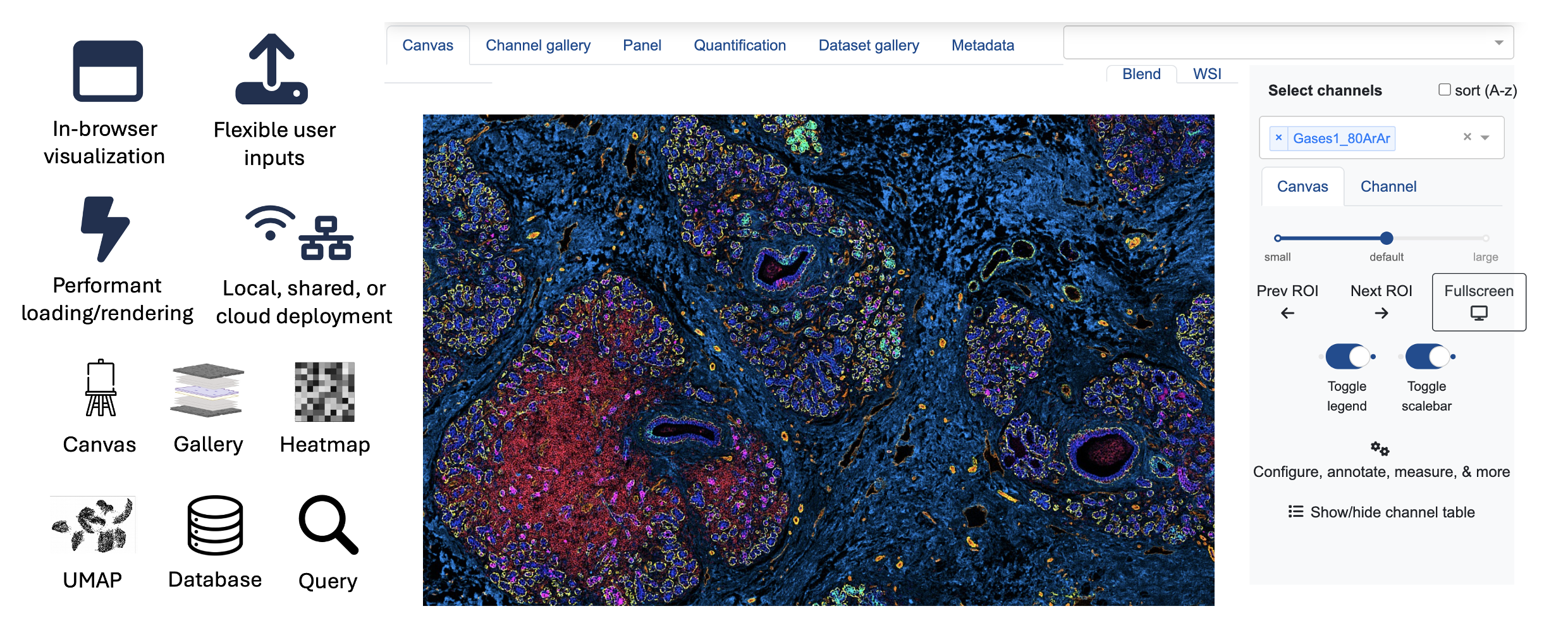
We develop computational tools and quantitative analytical approaches to facilitate the exploration and interpretation of complex spatial biology datasets. For example, we developed Rakaia (GitHub Page), an interactive, browser-based platform designed to make sophisticated spatial analysis tools accessible through an intuitive, user-friendly interface. By combining interactive data visualization, cellular annotation, and feature-based querying with advanced analytical capabilities, Rakaia enables researchers to efficiently explore, interrogate, and prioritize features of interest across large-scale multiplexed imaging datasets.
With thanks to all of our funding sources for their generous support of our work.


Publications
Latest Publications
Multimodal cell communication networks nominate immunotherapies for RCC subgroups with discrete T cell recruitment or expansion
Pfeil et al. bioRxiv. (2026)
Read Publication
Rakaia: interactive discovery of spatial biology at scale
An interactive platform for visualization, annotation, and analysis of highly multiplexed tissue imaging data.
Watson et al. bioRxiv. (2025)
Read Publication

Integrated spatial proteomics of human PDAC uncovers an expanded tumour-immune-stroma spectrum with genomic associations
Shakfa et al. bioRxiv. (2025)
Read Publication
Featured Publications
The single-cell pathology landscape of breast cancer
Jackson et al. Nature. (2020)
Read Publication

Imaging mass cytometry and multiplatform genomics define the phenogenomic landscape of breast cancer
Ali et al. Nat Cancer. (2020)
Read Publication
All Publications
All publications can be found on
Google Scholar: https://scholar.google.ca/citations?user=lS9OasAAAAAJ&hl=en
NCBI: https://www.ncbi.nlm.nih.gov/myncbi/hartland.jackson.1/bibliography/public/
Pubmed: https://pubmed.ncbi.nlm.nih.gov/?term=jackson+hw&sort=date
The People Behind the Science
Our Team

Hartland Jackson, PhD
Principal Investigator
hjackson@lunenfeld.ca

Beth Sunnucks
Lab Manager
sunnucks@lunenfeld.ca

Katie Teng
Graduate Student
kteng@lunenfeld.ca
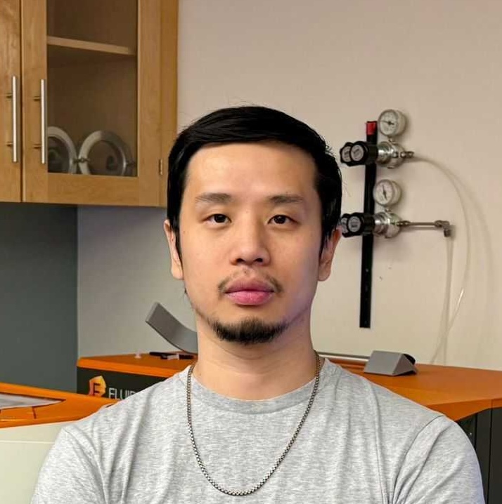
Eddy Chen, PhD
Post Doctoral Fellow
echen@lunenfeld.ca

Noor Shakfa, PhD
Post Doctoral Fellow
nshakfa@lunenfeld.ca
Sibyl Drissler
Graduate Student
sdrissler@lunenfeld.ca
Asma Amamou, PhD
Post Doctoral Fellow

Jennifer Gorman, PhD
Research Manager and
Scientific Associate
gorman@lunenfeld.ca

Matt Watson
Computational Scientist
mwatson@lunenfeld.ca

Ching Yeung Lam
Graduate Student
chingyeung@lunenfeld.ca

Darren Chan
Graduate Student
dchan@lunenfeld.ca

Tiak Tan
Graduate Student
tiaktan@lunenfeld.ca

Isabelle Hébert-Milette, PhD
Post Doctoral Fellow
hebert-milette@lunenfeld.ca

Alec Luchak, PhD
Post Doctoral Fellow

Avi DasGupta
Graduate Student
Pearl Sequeira
Research Administration
sequeira@lunenfeld.ca

Ferris Nowlan
Graduate Student
fnowlan@lunenfeld.ca
Golnaz Abazari
Graduate Student
gabazari@lunenfeld.ca

Jennifer Pfeil
Graduate Student
jpfeil@lunenfeld.ca

Matthew Perrone, PhD
Post Doctoral Fellow
mperrone@lunenfeld.ca

Yufei Zhang
Graduate Student

Ezi Ejiofor
Graduate Student
Our Alumni
Simon Latour, PhD
Post Doctoral Fellow
Taylor Larsen
Summer Research Student
Deni van Schie
Graduate Student
Anuroopa Dinesh
Lab Manager and
Research Technician
Ricardo Gonzalez, MD, PhD
Pathologist
Keiran Pace
Summer Research Student
Ari Forman
Graduate Student
Phoebe Ji
Summer Research Student
Somi Afiuni, PhD
Computational Scientist
Kellyona Khounborind
Summer Research Student
Naila Abid
Graduate Student
Lucy Colley
Technician
Scientific Collaborations
We are a highly collaborative team with many partnerships within the institute as well as with other labs across Toronto and beyond.
- We are developing new analysis methods for image cytometry and highly multiplexed imaging with the Campbell Lab at the Lunenfeld Tanenbaum Research Institute. https://www.camlab.ca/
- With the neighbouring Schramek Lab at LTRI we have created mouse model systems for the functional genomics study of single cells and multi-cellular tissues. https://schramek.lunenfeld.ca/
- Alongside the Notta Lab and the PanCuRx Initiative within the Ontario Institute for Cancer Research, we are investigating the single cell content of pancreatic ductal adenocarcinoma using a combination of ‘omics’ profiling methods. https://www.nottalab.com/
- In collaboration with the McGuigan Lab in BioEngineering at the University of Toronto, we are engineering multi-cellular tissue environments at scale. http://www.mcguiganlab.com/
- Working to develop new reagents for CyTOF and Imaging Mass Cytometry we work closely with the Nitz Lab in the Department of Chemistry at the University of Toronto. https://sites.chem.utoronto.ca/nitz/
Life in the Jackson Lab
Inside the Lab


Beyond the Bench


Join Our Team
Even though our lab is currently close to capacity we are still always looking for creative and enthusiastic scientists to join our team. In particular, we are looking to recruit talented post-doctoral fellows who believe they would have a unique skill set to contribute to our lab. If you have scientific research goals that you believe will align with ours, please contact Dr. Hartland Jackson directly to discuss your work and future interests.
Connect With Us
We are located at:
Lunenfeld-Tanenbaum Research Institute
Mount Sinai Hospital
Joseph & Wolf Lebovic Health Complex
Room 881 – 600 University Avenue
Toronto, Ontario Canada M5G 1X5
(416) 586-4800 x 8267